Pelvic Anatomy Dog - 3 - The pelvis is firmly attached to the spine (sacroiliac joint) and the limb is longer and more angulated than the thoracic limb (which is designed to bear weight and absorb impact).
byAdmin-
0
Pelvic Anatomy Dog - 3 - The pelvis is firmly attached to the spine (sacroiliac joint) and the limb is longer and more angulated than the thoracic limb (which is designed to bear weight and absorb impact).. The canine pelvis is relatively small and narrow. The pelvic limb is designed for propulsion. The canine pelvis is positioned between the dorsal and transverse planes and closer to the dorsal plane. The canine pelvis shape from a ventral view resembles a rectangle. Has cranial and caudal bellies in the dog only.
The canine pelvis shape from a ventral view resembles a rectangle. The hindlimb skeleton of the canine includes the pelvic girdle, consisting of the fused ilium, ischium, and pubis, and the bones of the hindlimb. Anatomy of the dog illustrated atlas this modules of vet anatomy provides a basic foundation in animal anatomy for students of veterinary medicine. It provides information about a dog's skeletal, reproductive, internal, and external anatomy, along with accompanying labeled diagrams. College of veterinary medicine • copyright 2013
Labeled Atlas Of Anatomy Illustrations Of The Dog from www.imaios.com With the large range of breeds and dog sizes, despite their difference in appearance, it might be surprising to hear dog anatomy is generally the same with regards to physical anatomy and characteristics. The size of hindlimb bones varies due to the significant variation in size for. Not visible on survey films because of their radiolucency and small size. However, dogs don't have a collar bone, unlike humans; Anatomy of the dog illustrated atlas this modules of vet anatomy provides a basic foundation in animal anatomy for students of veterinary medicine. That is exactly what you will find in this dogappy article. Not visible on survey films. The iliac crest is fused with the wing of the ilium in most dogs by 2 years of age, but a permanently incomplete fusion is common, occurring in more than 10% of dogs 3 ().this incomplete fusion can be misinterpreted easily as a fracture.
The muscles and nerves of the canine pelvic limb are reviewed including muscle actions.
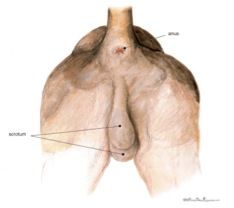
Dog anatomy is not very difficult to understand if a labeled diagram is present to provide a graphic illustration of the same. Urogenital system of the dog the pictures in this section are reprinted with permission by the copyright owner, hill's pet nutrition , from the atlas of veterinary clinical anatomy. The institute of canine biology. Über 7 millionen englischsprachige bücher. That is exactly what you will find in this dogappy article. The canine pelvis is positioned between the dorsal and transverse planes and closer to the dorsal plane. This veterinary anatomical atlas includes selected labeling structures to help student to understand and discover animal anatomy (skeleton, bones, muscles, joints, viscera, respiratory system. The pelvic girdle is formed by two hip bones which are joined ventrally at the cartilagenous pelvic symphysis and articulate dorsally with the sacrum. In dog compared the anatomical homologies of the perineal region in male and female dogs. Not visible on survey films because of their radiolucency and small size. The pelvic limb is designed for propulsion. College of veterinary medicine • copyright 2013 Assessment and management of pelvic fractures in dogs and cats (proceedings) march 31, 2009.
Not visible on survey films because of their radiolucency and small size. It has the ability to flex extend rotate adduct and abduct its whole limb because of this. The pelvic girdle is formed by two hip bones which are joined ventrally at the cartilagenous pelvic symphysis and articulate dorsally with the sacrum. Urogenital system of the dog the pictures in this section are reprinted with permission by the copyright owner, hill's pet nutrition , from the atlas of veterinary clinical anatomy. Secure the dog's pelvic limbs in this position using tape around the femurs at the level of the stifle joint (figures 2a and 2b).
Scielo Brasil 3d Printing Of Canine Hip Dysplasia Anatomic Models And Radiographs 3d Printing Of Canine Hip Dysplasia Anatomic Models And Radiographs from minio.scielo.br By means of roentgenological and morphological methods in 100% of cases, lateral, iliac and hypogastric ln are revealed. The acetabulum provides the socket to the joint of the hip and is composed of all three bones of the pelvis. The bone that articulates with the hip bones to form the hip joint is the femur. Not visible on survey films because of their radiolucency and small size. College of veterinary medicine • copyright 2013 Muscles of the pelvic limb; A non articular depression portion of the acetabulum used for the attachment of the ligament of the head of the femur. Anatomy of the dog illustrated atlas this modules of vet anatomy provides a basic foundation in animal anatomy for students of veterinary medicine.
K eep reading to learn more!.
The pelvic girdle is formed by two hip bones which are joined ventrally at the cartilagenous pelvic symphysis and articulate dorsally with the sacrum. The canine ischiatic or ischial tuberosities are wide and project caudally to form a broad ischiatic table. In 94% sacral and in 87.3% medial il … Not visible on survey films because of their radiolucency and small size. College of veterinary medicine • copyright 2013 The institute of canine biology. A non articular depression portion of the acetabulum used for the attachment of the ligament of the head of the femur. The hindlimb skeleton of the canine includes the pelvic girdle, consisting of the fused ilium, ischium, and pubis, and the bones of the hindlimb. The size of hindlimb bones varies a great deal because of the great variation in size for breeds of dogs. Muscles of the pelvic limb. A non articular depression portion of the acetabulum used for the attachment of the ligament of the head of the femur. The hindlimbs bear 40 of the dogs weight. They are most commonly seen in young, healthy dogs and cats.
In 243 mongrel female dogs anatomy, topography of the pelvic lymph nodes (ln), composition and frequency of their revealing have been studied. With the large range of breeds and dog sizes, despite their difference in appearance, it might be surprising to hear dog anatomy is generally the same with regards to physical anatomy and characteristics. By means of roentgenological and morphological methods in 100% of cases, lateral, iliac and hypogastric ln are revealed. They are most commonly seen in young, healthy dogs and cats. The canine pelvis is positioned between the dorsal and transverse planes and closer to the dorsal plane.
Leslie Gross Anatomy Pelvic Cavity Flashcards Cram Com from images.cram.com When properly aligned, the patella for each pelvic limb will be centered within the trochlear groove over the distal femur. Not visible on survey films. You will see that there are both ligaments and muscles that play important roles in stabilizing the hip joint, and developing the strength in these muscles through appropriate exercise is important for a. The acetabulum provides the socket to the joint of the hip and is composed of all three bones of the pelvis. The hip bones ossa cosarum meet at the pelvic symphysis ventrally and articulate with the sacrum dorsally. With the large range of breeds and dog sizes, despite their difference in appearance, it might be surprising to hear dog anatomy is generally the same with regards to physical anatomy and characteristics. The size of hindlimb bones varies a great deal because of the great variation in size for breeds of dogs. Medial aspect of the genual region.
It has the ability to flex extend rotate adduct and abduct its whole limb because of this.
In dog compared the anatomical homologies of the perineal region in male and female dogs. The hindlimb skeleton of the canine includes the pelvic girdle, consisting of the fused ilium, ischium, and pubis, and the bones of the hindlimb. A non articular depression portion of the acetabulum used for the attachment of the ligament of the head of the femur. The institute of canine biology: When properly aligned, the patella for each pelvic limb will be centered within the trochlear groove over the distal femur. Secure the dog's pelvic limbs in this position using tape around the femurs at the level of the stifle joint (figures 2a and 2b). The canine pelvis is relatively small and narrow. Muscles of the pelvic limb. Not visible on survey films. In 94% sacral and in 87.3% medial il … The acetabulum provides the socket to the joint of the hip and is composed of all three bones of the pelvis. By means of roentgenological and morphological methods in 100% of cases, lateral, iliac and hypogastric ln are revealed. The three components of each hip bone are the ilium, pubis and ischium.
A non articular depression portion of the acetabulum used for the attachment of the ligament of the head of the femur pelvic anatomy. It provides information about a dog's skeletal, reproductive, internal, and external anatomy, along with accompanying labeled diagrams.